Naason Science
Naason Science
  • Home
  • CRO Services
    • Preclinical Research
    • Histology Services
    • CNS Models
    • Pain Models
    • Cardiovascular Models
    • Ophthalmology Models
    • Internal Disease Models
    • Psychiatric Models
    • Bone and Autoimmune
    • Safety and Toxicity
    • In Vitro
  • News
  • Virtual Gallery
  • Investors
  • Contact
  • More
    • Home
    • CRO Services
      • Preclinical Research
      • Histology Services
      • CNS Models
      • Pain Models
      • Cardiovascular Models
      • Ophthalmology Models
      • Internal Disease Models
      • Psychiatric Models
      • Bone and Autoimmune
      • Safety and Toxicity
      • In Vitro
    • News
    • Virtual Gallery
    • Investors
    • Contact
  • Sign In

  • My Account
  • Signed in as:

  • filler@godaddy.com


  • My Account
  • Sign out

Signed in as:

filler@godaddy.com

  • Home
  • CRO Services
    • Preclinical Research
    • Histology Services
    • CNS Models
    • Pain Models
    • Cardiovascular Models
    • Ophthalmology Models
    • Internal Disease Models
    • Psychiatric Models
    • Bone and Autoimmune
    • Safety and Toxicity
    • In Vitro
  • News
  • Virtual Gallery
  • Investors
  • Contact

Account


  • My Account
  • Sign out


  • Sign In
  • My Account

Ophthalmology Modelling Platform

Naason Science provides advanced preclinical ophthalmology models to support research into a wide range of eye diseases and conditions. Our models are designed to replicate key pathological features of ocular disorders, enabling detailed studies on disease mechanisms and the evaluation of innovative therapeutic approaches. From glaucoma and dry eye disease to retinal degeneration and beyond, Naason’s ophthalmology models offer reliable and precise platforms for advancing ophthalmic research and developing impactful treatments. 

Rodent model of dry eye disease by chemical or surgical ELG excision

Dry eye disease is a common condition that affects the eye and causes symptoms including irritation, dryness, and blurred vision. Progression of dry eye can be evaluated by observation of the cornea by corneal fluorescence staining and tear volume measurement using the phenol red thread test.  


1. Rat model of dry eye disease by surgical excision of ELG 

Surgical removal of the extraorbital lachrymal gland (ELG) in rats severely limits tear secretion and induces symptoms of dry eye disease. 


2. Mouse model of dry eye by scopolamine and desiccating condition 

In this model, dry eye disease is induced by repeated injection with scopolamine. Dry eye stress is further induced by exposure to continuous airflow in a low-humidity environment. 

Mouse Models of Age-related Macular Degeneration (AMD)

Age-related Macular Degeneration (AMD) is a medical condition which may result in blurred or loss of vision in the center of the visual field due to damage to the macula of the retina and is one of the leading causes of vision loss and blindness in people over 50 years of age in developed countries. There are two forms of AMD; dry and wet AMD. 

NaIO3-induced dry AMD

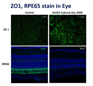
Naason Science offers a mouse model of dry AMD induced by NaIO3 injection. This model provides rapid phenotypic changes such as damage of tight junction integrity and thinning of retinal layers that can be assessed by immunohistochemical analyses. 

Laser-induced CNV model of wet AMD

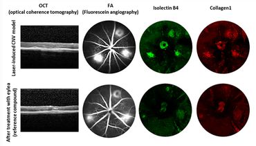
Dry AMD can progress to wet AMD when abnormal blood vessels develop underneath the retina. Wet AMD can be induced by photocoagulation of the retina using the 532 nm laser. The choroidal neovascularization (CNV) formed by the laser can be measured in vivo using optical coherence tomography and fluorescein angiography. We also offer immunohistochemical analyses on flatmounts using markers for CNV and subretinal fibrosis, which often accompanies wet AMD. 

Two-stage laser-induced Model of Subretinal Fibrosis

In this Model, the 532 nm laser is applied in two stages in the same lesion to produce a more severe phenotype of AMD and to induce subretinal fibrosis. Fibrosis is quantified by immunohistochemical staining after flatmounting. In this Model, the area of fibrosis increases 30 days after the second laser burn, providing a platform to test therapeutic efficacy of compounds.  

For more information please

Contact us

© 2017-2025 Naason Science Inc. All rights reserved.

  • Home
  • Preclinical Research
  • News
  • Areas of Expertise
  • Privacy & Terms
  • Contact

Powered by

Welcome to Naason Science website!

Our website uses cookies. By continuing to use this site, you accept our use of cookies.

DeclineAccept