Signed in as:
filler@godaddy.com
Naason Science offers a wide range of in vitro models designed to accelerate drug discovery and deepen understanding of disease mechanisms. Our in vitro platforms provide precise, controlled environments to study cellular processes, evaluate therapeutic efficacy, and identify potential drug targets. From cell-based assays to high-throughput screening systems, our models cover diverse research areas, including oncology, metabolic diseases, neurodegenerative disorders, and toxicology. With reliable and reproducible results, Naason Science’s in vitro models are an essential tool for bridging the gap between early-stage research and clinical development.

The primary cells of the CNS are divided into neurons and glial cells. Neurons are the main signaling cells of the nervous system, and each neuron can form connections with other neuronal cells through synapses.
Glia provides essential functions of neurons (nutrient provision, regulation of ion concentration, mediating immune response…) in various ways, and glial cell types include astrocytes, microglia, and oligodendrocytes.
Primary neurons and glia are used in many fields of neurological research, focusing on cellular mechanisms of the central and peripheral nervous system and neurodegenerative diseases.
Assay Analysis

Bone is in a constant state of remodeling, which is important for the maintenance of normal skeletal structure and function. Osteoclasts are responsible for aged bone resorption and osteoblasts are responsible for new bone formation. The resorption and formation is in stable at physiological conditions. However, when the balance is disturbed, bone architecture or function will be abnormal. Bone metabolism diseases, such as osteoporosis or osteopetrosis will occur.
Osteoblast – MC3T3 subclone 4
The MC3T3-E1 cell line established from newborn mouse calvaria is a well-known in vitro osteogenic model system and has been widely used in bone tissue engineering-related research. MC3T3-E1 cells display a sequential development pattern of proliferation and differentiation, resulting in calcified bone tissue similar to in vivo bone formation. The cells differentiate in accordance with the gene activation of osteoblast markers such as alkaline phosphatase (ALP), type I collagen and collagenase-1 (MMP-1), osteonectin (OPN), osteocalcin (OCN).
Assay Analysis

Osteoclast are multinucleated bone resorbing cells formed by cytoplasmic fusion of their mononuclear precursors. The Bone marrow-derived macrophages (BMDM) are primary macrophages obtained by in vitro differentiation of bone marrow cells in the presence of macrophage colony-stimulating factor (M-CSF or CSF1) and RANKL.
Assay Analysis:

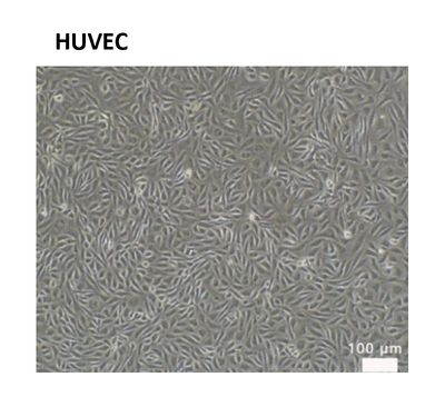
Angiogenesis, the formation of new vessels from pre-existing endothelium, is an important process in the adult organism because it supports the increasing demands for metabolic supplies (nutrients, various growth factors, and molecular oxygen) at sites of tissue repair or regeneration, during processes such as pregnancy, the female reproductive cycle, wound healing, and revascularization of ischemic tissue. Human umbilical vein endothelial cell (HUVEC) is used to study the function and pathology of endothelial cells. In particular, HUVEC have been used to study VEGF dependent angiogenesis. VEGF promotes the proliferation and migration of endothelial cells, stimulate the vascular permeability.
Assay Analysis
Cell proliferation
Migration
Tube formation:
Gene expression:
Protein expression: Western blot, ELISA
The in vitro lipid accumulation model using HepG2 cells is a valuable tool for studying non-alcoholic fatty liver disease (NAFLD) and related metabolic disorders. HepG2 cells, derived from human liver carcinoma, serve as a reliable platform for investigating lipid metabolism and the molecular mechanisms underlying steatosis. By inducing lipid accumulation in these cells, researchers can evaluate the efficacy of therapeutic compounds, explore pathways involved in lipid homeostasis, and identify potential targets for drug development. This model provides a cost-effective and scalable approach to advancing our understanding of liver-related metabolic diseases.


Assay Analysis